
 |
High-dimensionality Pattern Regression: Let Baseline Scans Tell More about Disease Progression
|
||
|
Many pathologies and diseases present a continuous spectrum of structural and functional changes. Therefore, it is important to discorver the potential relationship between brain changes and continuous clinical measures. A general high-dimensionaltiy pattern regression methodology was proposed, which was data-driven, and could be applied to many studies. The proposed method can predict continuous clinical progression from baseline scans. Meanwhile, it also provides diagnostic and prognostic indicators to identify neurodegenerative disease from imaging, potentially many years before clinical symptoms become measurable.
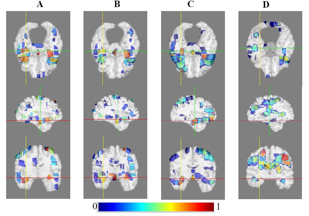
To reduce the huge dimensionality of original images while keeping sufficient information, an adaptive feature extraction method was proposed to generate spatial patterns, in which a measure of correlation between morphological measurements (voxel intensity) and a continuous clinical variable (such as MMSE) was used to group brain voxels into regions; Further feature selection was achieved using Relevance Vector Regression-Recursive Feature Elimination (RVR-RFE) under the bagging framework. Nonlinear RVM was then built to characterize the relationship between baseline scans and clinical variables. Applications:
Prediction of continuous disease progression;
Diagnostic predictors for early marker of AD;
Evaluation of cognitive performance;
1. Ying Wang, Yong Fan, Priyanka Bhatt, and Christos Davatzikos, "High-Dimensional Pattern Regression Using Machine Learning: From Medical Images to Continuous Clinical Variables ", NeuroImage, Volume 50, Issue 4, Pages 1519-1535, May 2010.
Software available soon! |
 |
Spatio-temporal Analysis of Individual Brain Changes: From MRI Image Series to Changing Brain Patterns to Clinical Stages |
||
|
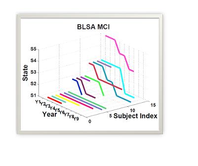
Brain changes due to development and maturation, normal aging or disease development are continuous, gradual, as well as various across individuals. By repeatedly evaluating each subject over time, longitudinal dataset are expected to provide more information for capturing the subtle brain changes.
Different from the traditional longitudinal study with group analysis, the HMM-based spatio-temporal analysis of regional brain pattern sequence on individuals was adopted. The proposed method was validated by medical images from two incomplete datasets (the ADNI and BLSA studies). State paths from the HMMs show the pathological brain changes during the observation period on an individual basis. By statistically analyzing the state paths and number of state transitions, an indicator to identify the subjects with a risk of MCI or AD was also provided. Applications:
Description of longitudinal development of elderly people; Predictive markers for early MCI and AD;
1. Ying Wang, Susan M. Resnick, and Christos Davatzikos, "Spatio-temporal Analysis of Brain MRI Images using Hidden Markov Models ", MICCAI, Volume 6362, Pages 160-168, 2010, Beijing . 2. Ying Wang, Susan M. Resnick, and Christos Davatzikos, " Analysis of Spatio-temporal Brain Imaging Patterns by Hidden Markov Models and Serial MRI Images ", to be submitted to NeuroImage soon. |
 |
Brain Patterns from Multi-Modality Images related to Specific Cognitive Domains
|
||
|
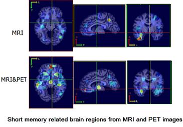
Human cognitive performance is quite complex, which links to specific brain areas with specific executive functions and memory processes. To gain a better understanding of this relationship, I developed a multi-parametric regression methodology by integrating the MRI and Positron Emission Tomography (PET e1) images, to detect brain regions associated with given cognitive domain.
Experimental results showed this method could detect the sensitive and specific brain regions related with the given cognitive functions. In addition, the regions found from the multi-modality measures were consistent with those from uniform modality. Especially for the short-term memory related brain regions, more significant brain areas were found from PET modality, which were different from the long-term memory case, where structure MRI contributes more to regression analysis.
1. Ying Wang, working with Joshua Goh, Susan M. Resnick, and Christos Davatzikos, "Joint Detection of Brain Regions with Specific Cognitive Comains: a Multi-parametric Regression Study ", |
|||
| Human Activity Recognition Based on R Transform
|
|||
|
Recognizing human activities from videos is a hot topic of research in computer vision, as the key technology to a wide range of applications such as intelligent surveillance, analysis of the physical condition of people and caring of aged people. The R transform as a shape descriptor to represent the activity was applied, and HMM employed to model the variations with time.
R transform has low computational complexity and geometric invariance. Compared with other commonly-used feature descriptors, R transform is robust to frame loss in video, disjoint silhouettes and holes in the shape, and thus achieves better performance in recognizing similar activities.
1. Ying Wang, Kaiqi Huang, Tieniu Tan, "Abnormal Activity Recognition in Office Based on R Transform", ICIP, 2007 2. Ying Wang, Kaiqi Huang, Tieniu Tan, "Human Activity Recognition Based on R Transform", CVPR, 2007 |
|||