

Multiple Sclerosis
Contact
People
- Christos Davatzikos, Kayhan Batmanghelich, Clyde Markowitz
Multiple Sclerosis (MS) is a chronic inflammatory disease of the central nervous system that affects the whole brain, manifest visibly as multi-focal lesions and subtle abnormalities, outside of the lesions, in the MRI scans. Diagnosis of MS is primarily based on clinical criteria but has found a reliable complement in MRI that provides a visualization of the lesions, and valuable insights into the underlying pathological processes of MS, and in studying the progression of the disease and treatment effects. Although lesions are the most evident indicator of abnormality, brain tissue outside of the lesions, called Normal Appearing Brain Tissue (NABT), comprising both the normal appearing white matter (NAWM) and grey matter (NAGM), is also affected in MS. It is expected that a characterization of the extent and severity of this abnormality in NABT will provide additional understanding of the pathology and thus could potentially have high diagnostic and prognostic value. The problem of characterizing this tissue abnormality is rendered challenging by several factors: the underlying pathology of MS is a complex combination of inflammation, demyelination, axonal injury and gliosis, with regression and progression in pathology over time. In addition, different MRI modalities reflect different aspects of the pathology, with the relatively newer modalities of Diffusion Tensor Imaging (DTI) and Magnetization Transfer (MT) imaging, demonstrating greater pathological specificity than the conventional ones of PD/T2 and FLAIR. Therefore, a comprehensive characterization of pathology induced abnormality in NABT requires the development of a computational framework that combines the spatial information of the extent of abnormality with the temporal information of changes in tissue pathology, obtained from several MR modalities.
The development of such a comprehensive computational multi-parametric spatio-temporal profile of abnormality of brain tissue, studying its evolution over time and obtaining a composite measure of extent and severity of abnormality is achieved in two phases:
Phase 1 (Spatial Characterization of Tissue Abnormality): To develop a multi-parametric tissue profile of abnormality in NABT by combining information from the MR protocols of T1, T2, FLAIR, GAD-enhanced, Magnetization Transfer (MT) imaging and Diffusion Tensor Imaging (DTI). The profile will comprise of 1) a spatial Tissue Abnormality Map (TAM) that provides a voxel-wise characterization of the tissue abnormality of NABT, created by using multimodality features with pattern classification techniques and 2) a composite abnormality score (CAS) of extent and severity of abnormality computed from these maps. We will evaluate the clinical significance and level of sensitivity of these scores by correlating them against the clinical measure consisting of the neurological, neurocognitive and ophthalmologic scores.
Phase 2 (Temporal Evolution of Tissue Abnormality): To develop a framework for quantifying the temporal evolution of tissue abnormality in terms of extent and severity, by incorporating the multi-parametric imaging profile designed in Phase 1 into a regression scheme to create temporal Tissue Abnormality Maps. We will evaluate the longitudinal sensitivity of the composite abnormality measure by correlating them with the clinical scores of longitudinal MR scans in these MS patients.
We hypothesize that 1) the multi-parametric tissue abnormality profile that we propose will provide a more comprehensive characterization of the extent and severity of abnormality in NABT tissue than any single MR modality; 2) the composite abnormality score computed from the tissue abnormality maps will help phenotype the disease into mild, medium and severe depending on the level of tissue identified to be at risk, thereby determining the nature of the treatment; 3) following these scores temporally will help determine treatment efficacy, leading to better prognosis and helping in testing new drugs; and 4) the multi-parametric framework will identify the reproducible MR protocols and their optimal combination that can best characterize tissue abnormality in MS.
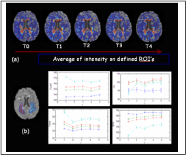
Temporal profile of abnormality and its variation in each of the MR protocols.
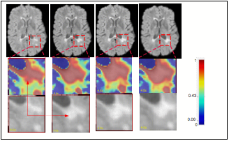
Probability map derived from two-class SVM classifiers: It shows the evolution of abnormality in a patient scanned 4 times. The top row shows slices from FLAIR images with a region of interest identified (indicated by the red dotted area) with a growing lesion. The inlaid color maps in the middle row are the visualization of the probabilistic abnormality map with the third row showing the growing lesion in the FLAIR image.
References
Nematollah Batmanghelich, Ragini Verma, “On non-linear characterization of tissue abnormality by constructing disease manifolds”, IEEE Computer Society Workshop on Mathematical Methods in Biomedical Image Analysis (MMBIA), Anchorage, Alaska, June 27 - 28, 2008.
Kayhan Batmanghelich, Xiaoying Wu, Clyde E. Markowitz, Ragini Verma, “Multiparametric MR analysis of temporal evolution of abnormality in MS”, ISMRM, Toronto, 3-9 May 2008.
Nematollah Batmanghelich, Xiaoying Wu, Clyde E. Markowitz, Ragini Verma, “Multiparametric Tissue Abnormality Characterization using Manifold Regularization”, SPIE Medical Imaging, San Diego, February 2008.
Ragini Verma and Peng Wang: “On Detecting Subtle Pathology via Tissue Clustering of Multi-parametric Data using Affinity Propagation” Mathematical Methods in Biomedical Image Analysis (ICCV), Brazil 2007.